#Dog lymph nodes skin
Histological image with hematoxylin and eosin staining of a cross section of a skin in the capillary-like network (scale bar: 5 mm) (left). Diagrams show changes of lymphatic pathways (bottom).

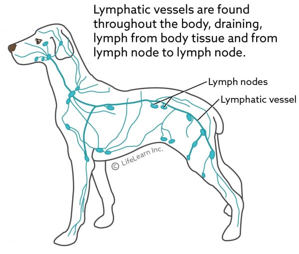
Lymphangiograms from the same dogs from lateral (left) and antero-posterior (right) views showing capillary-like network (black arrows) and bypassed lymph nodes (white arrows) (middle). Locations of lymph nodes are marked (white arrows). The ventral superficial lymph node (arrow) was identified using injections of ICG at interdigital webspaces.Ĭolor-coded diagram of the lymphatic territories (lymphosomes) with lymphatic vessels shown distally from their corresponding lymph nodes: 1, submandibular 2, parotid 3, dorsal superficial cervical 4, axillary 5, medial iliac 6, lateral sacral 7, hypogastric 8, popliteal 9, superficial inguinal 10, ventral superficial cervical.Ī montage of indocyanine green lymphographic images of the left forelimbs of 2 live dogs 6 months after lymph node dissection (top).īright spots were seen in the area in which the surgery took place (black arrow). Montage of indocyanine green (ICG) lymphographic images of the left forelimb of the live dog prior to lymph node dissection. The popliteal lymph node with afferent lymphatic vessels (stained orange by the lead tetroxide radiocontrast mixture).Īnteroposterior (top) and lateral (bottom) radiographs of the whole body carcass after lymphatic injection.


Marked lymphatic vessels near the dorsal midline in the torso (indocyanine green injection sites are shown as green dots) (top).Ī magnified photo shows that ICG was specifically taken into the lymph vessel (arrow) (bottom). Tracing of the lymphatic vessels visualized using ICG lymphography (right). Indocyanine green (ICG) fluorescent lymphographic image of the medial side of the left forelimb in a dog carcass (left).


 0 kommentar(er)
